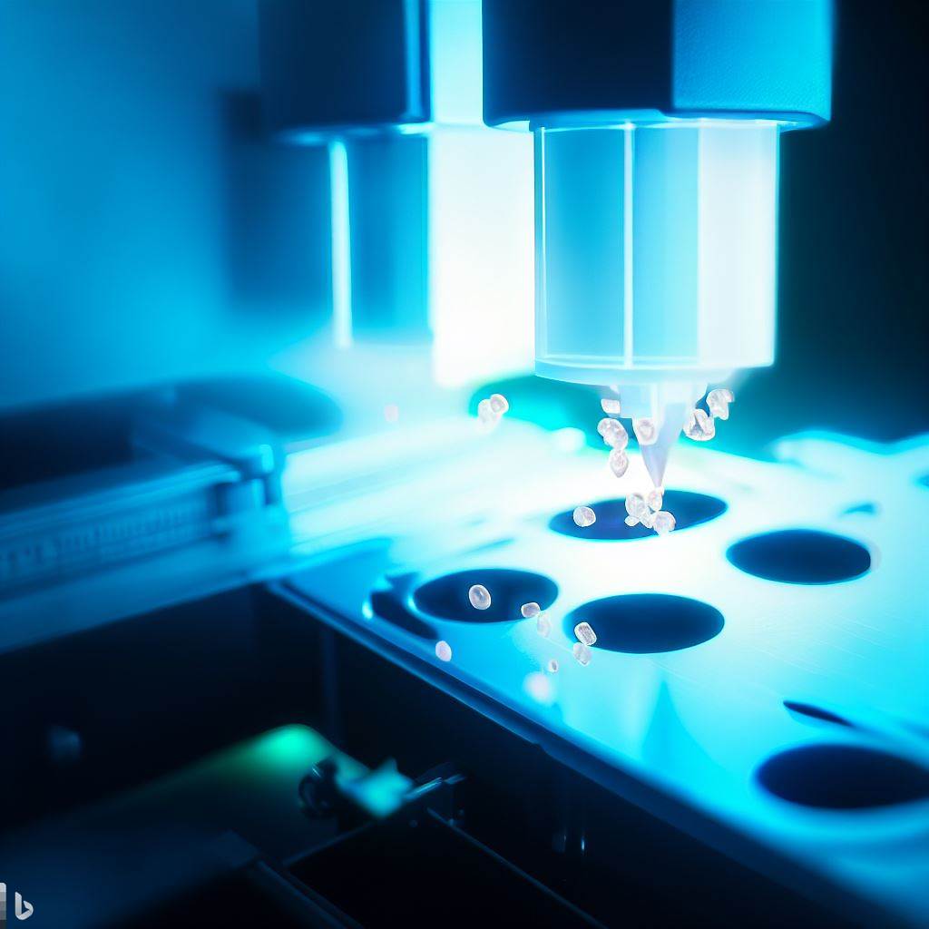
Blood flow cytometry is a technique that uses a device called a flow cytometer to measure and analyze the physical and chemical properties of blood cells. Blood flow cytometry can provide valuable information about the number, size, shape, and function of different types of blood cells, such as red blood cells, white blood cells, and platelets. Blood flow cytometry can also identify and quantify various molecules and markers that are present on or inside the blood cells, such as proteins, antibodies, antigens, DNA, and RNA. Blood flow cytometry can be used for various purposes, such as diagnosis, monitoring, research, and therapy of various diseases and conditions that affect the blood cells, such as anemia, infection, inflammation, leukemia, lymphoma, and others. In this text, I will explain how blood flow cytometry works, what are its advantages and disadvantages.

Working principle
Blood flow cytometry works by using the principles of fluidics, optics, and electronics. The process of blood flow cytometry consists of the following steps:
- Sample preparation. A blood sample is collected from a patient and treated with a special solution that prevents clotting and preserves the cells. The blood sample is then mixed with fluorescent dyes or antibodies that bind to specific molecules or markers on or inside the blood cells. These fluorescent dyes or antibodies emit light of different colors when they are excited by a laser beam. The fluorescent dyes or antibodies can be used to label different types of blood cells or different subtypes of the same type of blood cell.
- Flow cytometer. The flow cytometer is a device that consists of four main components: a fluidic system, an optical system, an electronic system, and a computer system. The fluidic system transports the blood sample into a narrow stream that passes through a laser beam. The optical system consists of one or more lasers that excite the fluorescent dyes or antibodies on the blood cells and one or more detectors that collect the light signals emitted by the blood cells. The electronic system converts the light signals into electrical signals and amplifies them. The computer system analyzes the electrical signals and displays the results in graphical and numerical formats.
- Result presentation. The results of blood flow cytometry are presented in two main formats: histograms and scatter plots. Histograms show the distribution of blood cells according to one parameter, such as cell size or fluorescence intensity. Scatter plots show the relationship between two parameters, such as cell size and fluorescence intensity. Each dot on a scatter plot represents one cell. The results can also be presented in other formats, such as tables or charts.
Advantages and Disadvantages of Blood Flow Cytometry
Blood flow cytometry has several advantages over other methods of blood cell analysis, such as:
- High sensitivity and specificity. Blood flow cytometry can detect and differentiate even rare or abnormal blood cells that may be missed by other methods. Blood flow cytometry can also identify and quantify various molecules and markers that are present on or inside the blood cells with high accuracy and precision.

- High speed and throughput. Blood flow cytometry can process and analyze thousands of blood cells per second, which is much faster than other methods that can take minutes or hours. Blood flow cytometry can also analyze multiple parameters of blood cells simultaneously, which can save time and resources.
- High versatility and flexibility. Blood flow cytometry can be used for various purposes, such as diagnosis, monitoring, research, and therapy of various diseases and conditions that affect the blood cells. Blood flow cytometry can also be adapted to different types of blood samples, such as whole blood, plasma, serum, or bone marrow.
- However, blood flow cytometry also has some disadvantages, such as:
- High cost and complexity. Blood flow cytometry requires expensive and sophisticated equipment, such as flow cytometers, lasers, detectors, computers, and software. Blood flow cytometry also requires skilled and trained personnel to operate and maintain the equipment, as well as to interpret and validate the results.
- High variability and standardization. Blood flow cytometry can be affected by various factors that can introduce variability and error in the results, such as sample quality, sample preparation, staining protocol, instrument settings, data analysis, and data interpretation. Blood flow cytometry also lacks universal standards and guidelines for quality control and quality assurance.
How AI image analysis complements flow cytometry
AI image analysis and flow cytometry can complement each other in various ways, such as:
- Increasing the accuracy and reliability of blood cell identification and classification. AI image analysis can use algorithms that are trained on large databases of blood cell images from different sources and conditions. These algorithms can recognize and classify different types of blood cells with high accuracy and consistency, regardless of the variations in staining quality, slide preparation, image quality, or operator skill. Flow cytometry can use algorithms that can measure various parameters of blood cells, such as hemoglobin concentration, cell volume, cell shape, cell surface markers, and cell internal structures. These parameters can help to differentiate blood cells that have similar appearance or fluorescence intensity.
- Providing more information and insights about blood cell morphology and function. AI image analysis can provide high-resolution and high-quality images of blood cells that can reveal more details and features than flow cytometry. AI image analysis can also detect any abnormal or immature cells that may indicate a disease or a condition. Flow cytometry can provide quantitative and objective data about blood cells that can reflect their function and status. Flow cytometry can also identify and quantify various molecules and markers that are present on or inside the blood cells, such as proteins, antibodies, antigens, DNA, and RNA.
- Enhancing the versatility and flexibility of blood cell analysis. AI image analysis and flow cytometry can be used for various purposes, such as diagnosis, monitoring, research, and therapy of various diseases and conditions that affect the blood cells. AI image analysis and flow cytometry can also be adapted to different types of blood samples, such as whole blood, plasma, serum, or bone marrow. AI image analysis and flow cytometry can also be integrated with other technologies, such as digital microscopy, optical microscopy, or molecular biology.
AI image analysis and flow cytometry are powerful techniques that can provide comprehensive and detailed information about blood cells and their changes. AI image analysis and flow cytometry can complement each other in many aspects and improve the quality and efficiency of blood cell analysis.