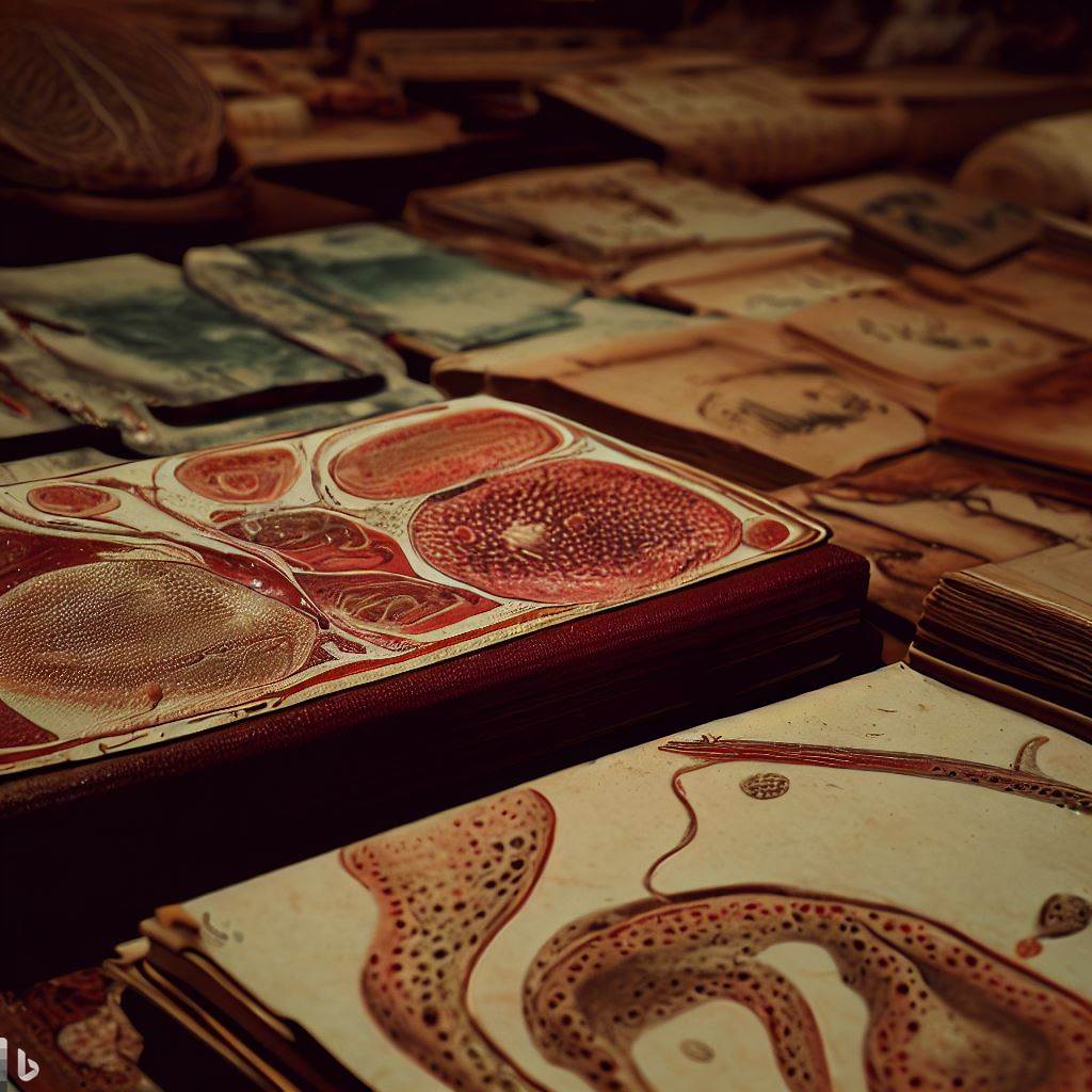
Histopathology and cytology are sciences of form that have largely depended on the study of the dead: dead bodies, dead tissues, and dead cells. Each science began with observers isolating, identifying, and naming the external and internal structures of living things, first with the naked eye and then with microscopes. For some investigators, the primary goal has been classification, arranging the bewildering array of plants, insects, fish, birds, and animals into groups and subgroups based on the shapes and arrangements of their parts.

The birth of science
The earliest phase of cytology began with the English scientist Robert Hooke’s microscopic investigations of cork in 1665. He observed dead cork cells and introduced the term “cell” to describe them. However, it was not until the 1830s that the German botanist Matthias Schleiden and the German zoologist Theodor Schwann proposed that all living organisms are composed of cells. This was the beginning of the cell theory, which states that cells are the basic units of life and that new cells arise from pre-existing cells.
Histopathology, on the other hand, emerged from the field of pathology, which is the study of diseases and their causes. The father of modern pathology is considered to be the French physician Marie François Xavier Bichat, who in 1801 published his Treatise on Membranes. He divided the human body into 21 systems of tissues, each with its own characteristics and functions. He also distinguished between normal and abnormal tissues, and suggested that diseases affect specific tissues rather than whole organs.
The development of histopathology was greatly influenced by the invention of new staining techniques that allowed different types of cells and tissues to be distinguished by their color under the microscope. One of the pioneers of staining was the German physician Rudolf Virchow, who in 1858 published his Cellular Pathology. He applied the cell theory to pathology and stated that diseases are caused by changes in cells. He also coined the term “biopsy” to describe the removal and examination of a small piece of tissue from a living person.
20th century
One of the major developments in histopathology in the 20th century was the improvement of staining techniques. Staining is the process of applying dyes or chemicals to tissue sections to enhance their contrast and visibility under the microscope. Staining can also reveal specific structures or molecules that are relevant for diagnosis or research. In the early 20th century, several new staining methods were introduced, such as the Gram stain for bacteria, the Ziehl-Neelsen stain for tuberculosis, the Giemsa stain for malaria, and the silver stain for nerve fibers. These stains enabled pathologists to identify and classify various microorganisms and diseases.
Another important breakthrough in histopathology in the 20th century was the discovery of immunohistochemistry (IHC). IHC is a technique that uses antibodies to detect antigens or proteins in tissue sections. Antibodies are molecules that bind to specific antigens with high specificity and affinity. By labeling antibodies with fluorescent or enzymatic markers, pathologists can visualize and quantify the expression of antigens in tissues. IHC was first developed in the 1940s by Albert Coons and colleagues, who used fluorescent antibodies to detect pneumococcal antigens in infected mice. Since then, IHC has become a powerful tool for diagnosing and studying various diseases, such as cancer, autoimmune disorders, infectious diseases, and neurological disorders.
A third major achievement in histopathology in the 20th century was the invention of electron microscopy (EM). EM is a technique that uses electrons instead of light to magnify and image specimens. EM can achieve much higher resolution and magnification than light microscopy, allowing pathologists to observe subcellular structures and organelles. EM was first developed in the 1930s by Ernst Ruska and Max Knoll, who built the first prototype of an electron microscope. In the 1950s and 1960s, EM was applied to histopathology by pioneers such as George Palade, Keith Porter, and Christian de Duve, who revealed the ultrastructure and function of cells and tissues. EM has contributed to the discovery of many cellular components and processes, such as ribosomes, lysosomes, endocytosis, and apoptosis.
And at the end of the 20th century, digital scanners began to appear.
Digital scanners
Digital scanners can improve histopathology in several ways, such as:
- Increasing the accessibility and scalability of histopathology. Digital scanners can enable remote consultation and collaboration among pathologists across different locations. They can also facilitate the integration of histopathology with other data sources, such as genomic, transcriptomic, or clinical data.
- Improving the workflow efficiency and productivity of pathologists. Digital scanners can reduce manual labor and errors in preparing, transporting, and storing glass slides. They can also allow faster and more accurate diagnosis and reporting of histopathology results.
- Enhancing the quality and scope of histopathological analysis. Digital scanners can provide better image quality and resolution than conventional microscopes. They can also enable the use of advanced image analysis techniques, such as artificial intelligence (AI), to assist in the interpretation and quantification of histopathology images.

Conclusion
In conclusion, histopathology has made significant progress and discoveries in the 20th century, thanks to the development of new techniques and technologies. Staining, immunohistochemistry, and electron microscopy have enhanced the quality and scope of histopathological examination and analysis. Histopathology has also become more integrated with other disciplines, such as molecular biology, genetics, immunology, and pharmacology. Histopathology continues to play a vital role in medicine and science in the 21st century.