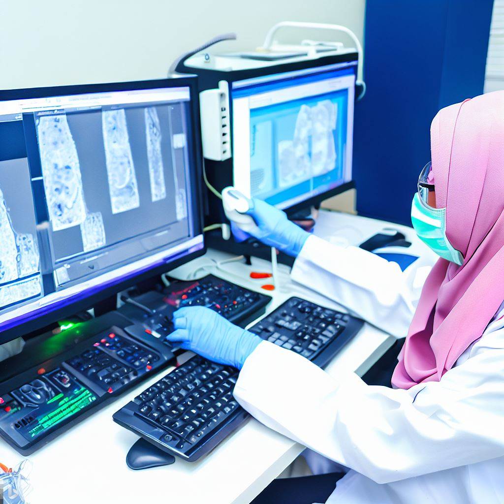
Digital microscopy is a method of microscopy that uses special cameras and computers to obtain, process and analyze images of microscopic objects. Unlike traditional optical microscopy, digital microscopy does not have eyepieces, but instead transmits the image to a screen or another output device. This makes working with digital microscopes more comfortable, fast and accurate. In this text, we will tell you about the main advantages of digital microscopy in medicine and the areas of its application.
Advantages of Digital Microscopy in Medicine
Digital microscopy has a number of advantages over traditional optical microscopy in medicine, such as:
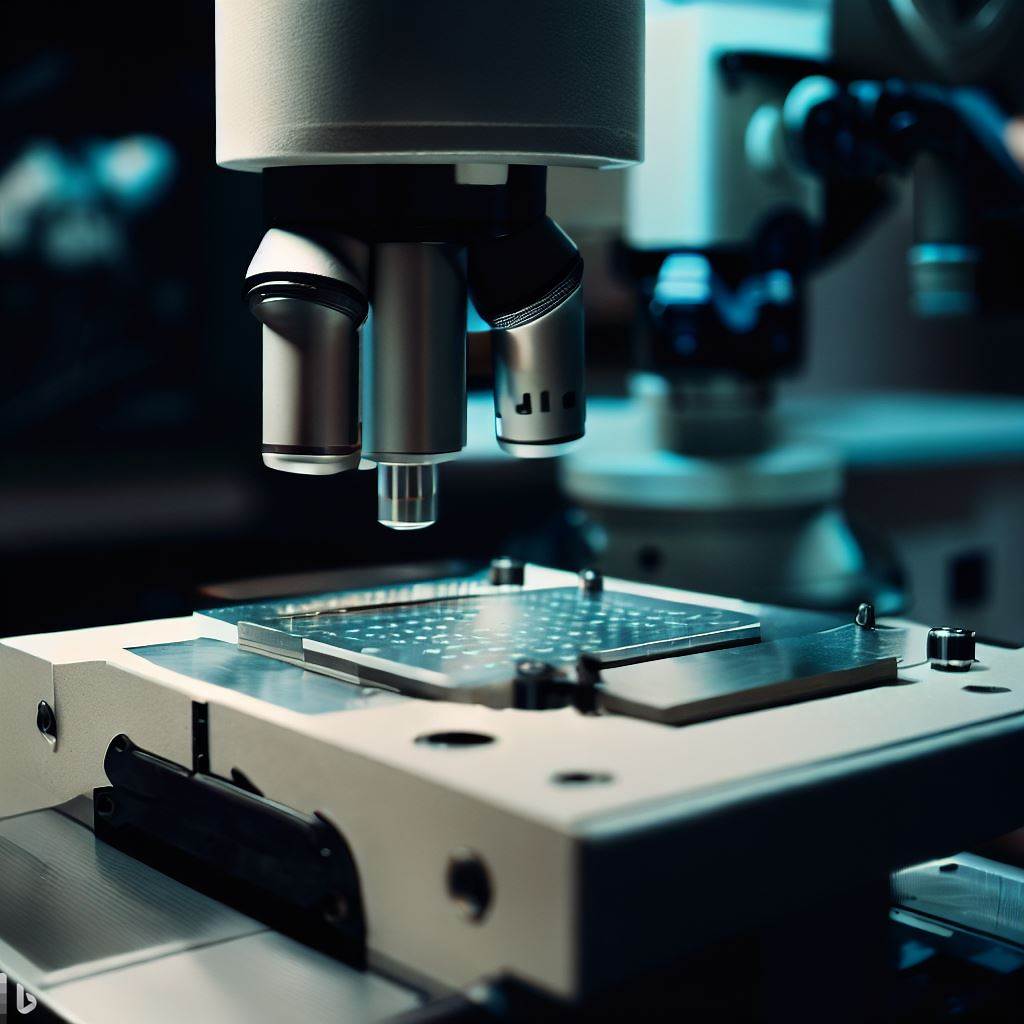
- High resolution images. Digital microscopes use cameras with a large number of pixels, which allow obtaining clear and detailed images of cells, tissues and organs. In addition, digital microscopes have the ability to change the angle of inclination and rotation of the camera, which gives the possibility to obtain two-dimensional and three-dimensional images of objects from different perspectives. This improves the quality of diagnosis and research, as it allows seeing the structure and function of objects more fully.
- Simplicity and convenience of use. Digital microscopes do not require setting up eyepieces, focusing and lighting, as all these parameters are adjusted automatically or with the help of special software. The user can observe the object on a large screen, without straining the eyes and neck. Also, digital microscopes allow saving multiple user profiles, which is convenient if one microscope is used by different specialists. Digital microscopes also save time and resources, as they do not require transportation and storage of glass slides with samples.
- Large possibilities of analysis and documentation. Digital microscopes allow not only obtaining images of objects, but also performing various measurements, annotations, comparisons, classifications and statistical analysis of data. All these functions are available in special software, which also allows saving, transferring and printing the obtained results . Digital microscopy also contributes to the development of deep learning, which uses neural networks for automatic recognition and interpretation of images.
Areas of Application of Digital Microscopy in Medicine
Digital microscopy has found wide application in various fields of medicine, such as:
- Diagnosis and treatment of diseases. Digital microscopy helps to determine the causes and characteristics of diseases, such as infections, cancer, anemia, allergy and others. With the help of digital microscopes, one can perform analysis of blood, urine, sperm, smears, biopsies and other biological materials. Digital microscopy also allows observing the dynamics of disease and effectiveness of treatment, as well as consulting with other specialists remotely.
- Research and development. Digital microscopy contributes to the development of science and technology in medicine, as it allows studying the properties of cells, tissues and organs at the micro level. With the help of digital microscopes, one can conduct experiments, testing, modeling and optimization of processes and products. For example, digital holographic microscopy allows obtaining three-dimensional images of living cells without staining and damaging them. This can be useful for studying cell cycle, apoptosis, differentiation and other processes.
- Education and training. Digital microscopy is an effective tool for teaching students and improving the skills of doctors. With the help of digital microscopes, one can demonstrate various objects and phenomena on a large screen or projector, as well as use interactive elements such as zooming.
Celly provides a low-cost automated microscope that allows you to move diagnostics into digital format.